Tick Identification and Information

Lyme disease is all about ticks. The more you know about ticks, the more you will understand Lyme disease and other illnesses caused by ticks, and the more knowledge you will have to prevent them.

Ticks are blood-feeding arthropods belonging to arachnid family, closely related to spiders and scorpions. With over 900 species identified worldwide, these small arachnids are remarkably adaptable, inhabiting a range of environments from dense forests to urban gardens. In the U.S., there are 15 species known to transmit a total of 30 human pathogens, but most of these are rare. Just three tick species are responsible for 98% of all tick-borne illness in the U.S.

Ecologically, ticks are important as they are part of the food chain and contribute to controlling animal populations. As ectoparasites, they feed on the blood of mammals, birds, reptiles, and occasionally amphibians. This feeding behavior can impact their hosts in various ways, ranging from minor irritation to serious health issues. Some species of ticks are highly specialized and feed on only specific host species, contributing to the delicate balance in natural ecosystems.

Every tick species has a complex life cycle which often involves multiple blood meals from different organisms, they have the ability to become vectors for disease-causing pathogens, including bacteria and viruses that are of importance to human public health. When they feed on their hosts, ticks can transmit pathogens such as bacteria and viruses.

This makes ticks notorious for their role in the spread of illnesses like Lyme disease, Rocky Mountain spotted fever, and several other infections. The incidence of some of these tick-borne diseases has been on the rise in the US due to a variety of factors such as climate change, increased land development, and other ecological factors. This underscores the importance of evidence-based information on how to prevent tick bites, remove ticks, and understand the science behind tick-borne illnesses.

The blacklegged tick, Ixodes scapularis, has two common names because there is disagreement among entomologists that it is a single species. The range of I. scapularis extends from Florida and Texas in the South to Maine and Wisconsin in the North, but there are both genetic and behavioral difference between the southern and northern forms. It was unknown in most of the northern states until Lyme disease was discovered and it was described as a new species, Ixodes dammini, the deer tick. The common name of deer tick is still widely used in the northern states where Lyme disease is prevalent. The blacklegged ticks in the southern states rarely bite humans and few are infected with Borrelia burgdorferi. Consequently, few cases of Lyme disease occur in that area. The following discussion is about deer ticks, the northern form of I. scapularis, although their basic biology is similar.

The deer tick has three feeding stages, larva, nymph and adult. Each stage feeds only once before changing into the next stage. Larvae and nymph attach and feed on blood for 3-4 days and the adult female feeds for 7 days. Adult males do not feed on blood. After feeding is completed, the tick falls off and digests the blood meal, which takes about a month before it changes into next stage. Engorged female ticks then lay eggs, about 2,000 of them, and then die. Male ticks die after mating with one or more females while they are feeding.

All of this takes place in wooded areas where animal hosts are abundant. The immature stages, larva, and nymph, of the deer tick feed upon small mammals and birds which can infect ticks with Borrelia burgdorferi, while the adult stage feeds primarily upon deer. Because mating and female feeding occur on deer, it is considered to be the reproductive host, hence the name deer tick.

The southern form also feeds upon deer in the adult stage, but larvae and nymphs frequently feed upon lizards, which are not infected with B. burgdorferi. Thus, the risk of getting Lyme disease in the South is very low compared to the northern states.

Each stage of the deer tick is actively seeking hosts at different times of the year. Larvae host-seek in August and nymphs host-seek from May to July. Adults seek hosts from late October until early May, depending upon the temperature. They cannot host-seek when the temperature is below 45 degrees or when there is snow on the ground.

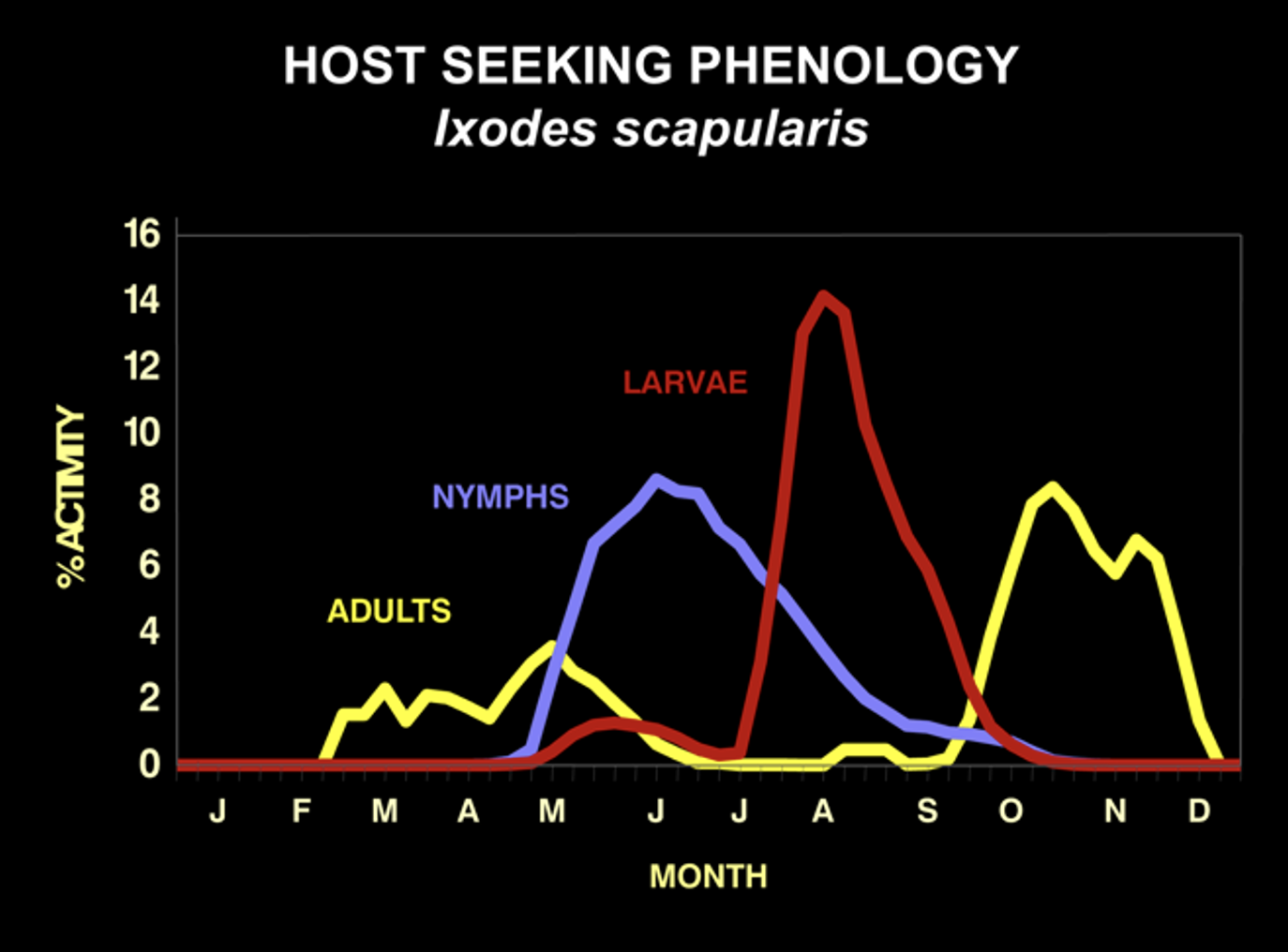
The seasonality of host-seeking has important epidemiological significance as the nymphal stage is responsible for 80 to 90% of Lyme disease cases. This is because they are small and hard to see, and they are active during the summer when outdoor activities are at their peak. They commonly feed to repletion (3-4 days) on people without notice. In fact, only about 25% of Lyme disease patients recall a tick bite. This is why knowing about deer tick biology is essential for Lyme disease prevention.

In endemic areas, about 20% of nymphs and 50% of adult deer ticks are infected with the Lyme disease spirochete Borrelia burgdorferi. Bites by adult ticks do not usually cause Lyme disease because they are larger and more easily noticed than nymphs. They are also active in months with less out-door activity. But, where there are adults, there must have been nymphs earlier in the summer.

Transmission of the Lyme disease spirochete occurs after 36 hours of feeding. Finding an attached tick and removing it before that time will prevent Lyme disease. Adult ticks are usually found and removed before that time, but not so for nymphs. That’s why routine tick checks are important for Lyme disease prevention. If you find an attached tick, there are ways to determine how long it has been feeding. The chart below estimates the rate of engorgement over time-based upon animal studies. An engorgement time greater than 36 hours is considered to be risky and prophylactic antibiotic therapy is recommended.

Nymphal Stage

Adult Female

Being bitten by a tick after that time does not mean that you will get Lyme disease. There is an 80% chance that the tick was not infected with B. burgdorferi and not everybody bitted by an infected tick will get Lyme disease. Only about 2% of patients who experience a tick bite will develop Lyme disease. Watch for the development of redness at the tick-bite site. A localized redness reaction to the tick bite is common, especially if you have been bitten before. But if it expands beyond 4 inches, seek immediate medical attention for prompt antibiotic therapy. It is important to note that other pathogens transmitted by deer ticks can infect at much earlier feeding times, minutes for Powassan virus and hours for anaplasmosis, and that all stages can be infected with Powassan virus or Borrelia miyamotoi.

Preventing tick bites is the first step in reducing the risk of contracting Lyme disease and other tick-borne illnesses. There are many ways to prevent tick bites. The most obvious way is to stay out of the woods, but that is not an option for most people. If you live in or visit areas where ticks are known to occur, you should take precautions. Your local health department may be able to advise you of tick-prone areas or local cases of tick-borne disease.  

  • Personal protection:
Stay on trails

When hiking, avoiding wooded and brushy areas, high grasses, and leaf litter. 

Wear light colored clothing

To make crawling ticks easier to see and remove. Tucking pants into socks will prevent ticks from crawling under clothing where they may be missed. Wearing closed-toe shoes over sandals when in high-risk areas, or even opting for knee-high rubber boots can prevent ticks from ascending as they have difficulty crawling on smooth surfaces.

Apply repellents to your skin

The use of repellents is highly recommended. Products containing DEET or Picaridin are effective for repelling ticks from exposed skin. Other repellents have not been consistently demonstrated to be effective at repelling ticks. For tick protection, you do not need to apply repellents to the face or arms as ticks crawl up your body from leaves or vegetation close to the ground.

DEET

Apply repellents that contain 20% or more DEET on exposed skin for long-lasting protection. Protection duration with 20% DEET can vary, but it typically provides 4 to 8 hours of protection against ticks. Factors such as temperature, humidity, sweating, and water exposure can influence how long the protection will last. If you’re spending a prolonged period outdoors, especially in areas where tick exposure is a concern, it is advisable to reapply the repellent according to the product’s instructions.


To maintain effective protection, users should follow the guidance provided on the repellent’s label regarding reapplication times. If you’re also using sunscreen, DEET should be applied after sunscreen, and keep in mind that reapplication will need to adhere to the most frequent interval recommended by either product. Higher DEET concentrations provide longer protection.

Picaridin

20% concentration of picaridin can provide up to 8-12 hours of protection against ticks. The exact duration can vary depending on several factors including the specific product formulation, the species of tick, and environmental conditions such as temperature, humidity, and exposure to water.


It is important to read and follow the product’s instructions for application and reapplication. If you are in an area with a high risk of tick bites, be sure to reapply the repellent as directed to maintain effectiveness.

Treat clothing and gear with permethrin

Permethrin is an insecticide that is highly effective at killing ticks that may crawl on you, especially if you are in areas with high tick activity. Treat clothing and gear, such as boots, pants, socks, and tents, with products containing 0.5% permethrin according to instructions. Permethrin can be protective through several washings.

Take a shower

Take a shower within two hours of coming indoors. This can wash away unattached ticks and provides a good opportunity to do a skin check.

After being outdoors, perform a thorough tick check

Check your clothing, gear, and pets for ticks. Ticks generally take between 15 minutes and 2 hours to crawl around and find a suitable place to attach and feed, so being prompt about checking for any unattached ticks can reduce your risk of potential tick bites. Conduct a full-body check upon return from potentially tick-infested areas, which includes under the arms, the groin/genital areas, in and around the ears, inside the belly button, behind the knees, between the legs, around the waist, and especially in the hair. Ticks are often attracted to warmer areas of the body, so make sure to check all creases and warm area folds. Since ticks can be very small, it’s important to feel for bumps on the skin that might not be immediately visible.

Children may not be as diligent about tick checks, so it’s important for adults to check them, paying close attention to their hair, ears, waist, and groin. If you were outdoors earlier in the day, do one more tick check before bed, especially if you’ve been in a high-risk area. Ticks can migrate for several hours before settling down to attach and feed.

Remove any unattached ticks that may be on pets

Remove any unattached ticks that may be on pets after having been outdoors. Consult with your veterinarian for recommended tick control products. Many of these require ticks to bite your bite, so it is important to thoroughly comb your pets after they’ve spent time outdoors if you live in an area with high tick activity.

Insecticides

Treating high-risk areas, such as residential lawns, especially those adjacent to woods, with an insecticide can reduce tick populations by up to 90%. Several insecticides are approved by the EPA for tick control. Commercial applicators can perform this service, but it can also be done by homeowners. Granular formulations penetrate the ground where ticks live without adhering to blades of grass where human exposure may occur. A single application in April or early May will kill nymphal ticks before they become active in June and July as they overwinter in the ground. A single application in September or early October will have the same effect for adult ticks which become active in late October and remain active until spring, depending upon temperature (above 45 degrees).

Bait boxes and tubes

There are several products on the market designed to prevent ticks from feeding upon mice and chipmunks by directly applying insecticides. This would also prevent ticks from becoming infected with Borrelia burgdorferi and other pathogens harbored by these animals. Studies have had mixed results and may result in a false sense of security since tick bites are frequently unnoticed making it difficult to evaluate their effectiveness. There is also a product on the market that is designed to vaccinate mice against Lyme disease with an oral bait administered by a bait box which shows some promise, but it has not been independently evaluated. It would not prevent ticks from acquiring other tick-borne pathogens.

Deer fencing and deer reduction

Deer are the reproductive host for I. scapularis and adult ticks do not occur or reproduce in the absence of deer. Deer exclusion by fencing must be done over large areas of several acres because nymphal ticks are disseminated by mice and other small animals. Deer reduction by professional marksmen can reduce local deer populations, but a threshold of deer density that would reduce the abundance of ticks sufficient to reduce Lyme disease risk has yet to be determined. Hunting deer is not an effective method for preventing tick-borne diseases because it does not significantly reduce the deer population and it is prohibited in most residential areas.

If you find a tick attached to your skin, it’s important to remove it as soon as possible to reduce the risk of disease transmission. Removal of black-legged ticks within 36 hours, even if the tick is infected with Borrelia burgdorferi, nearly eliminates your risk of contracting Lyme disease.

It is only recommended that you remove ticks mechanically using fine-tipped tweezers.

  • Pull skin taut to allow as much access to the tick mouthparts as possible.
  • Grasp the tick as close to the skin’s surface as possible. Avoid using blunt tweezers or your fingers, as this can increase the risk of tearing the tick and spreading any potential infection.
  • Pull Upwards with steady, even pressure. Don’t twist or jerk the tick, as this can cause parts of the tick to break off and remain in the skin. If this happens, remove the mouthparts with tweezers. If you are unable to remove the mouth easily with clean tweezers, leave it alone and let the skin heal.
  • Clean the bite area and your hands. After removing the tick, thoroughly clean the bite area and your hands with rubbing alcohol or soap and water.
  • Dispose of the tick. Submerse the live tick in alcohol, place it in a sealed bag/container, wrap it tightly in tape, or flush it down the toilet. Try to avoid crushing a tick with your fingers.
  • Avoid “Alternative” removal options. DO NOT use nail polish, petroleum jelly, or heat to make the tick detach from the skin. These methods are not effective and may increase the likelihood of transmitting a pathogen to the host or harm.

After tick removal, monitor the bite site over the next few weeks for any signs of Lyme disease or other tick-borne illnesses, such as a rash or fever. Not all ticks carry disease, and not all tick bites will result in disease. However, if you have concerns or symptoms develop, consult with a healthcare provider. They may advise you based on the type of tick, your geographical area, the duration of attachment, and the local prevalence of tick-borne illnesses.

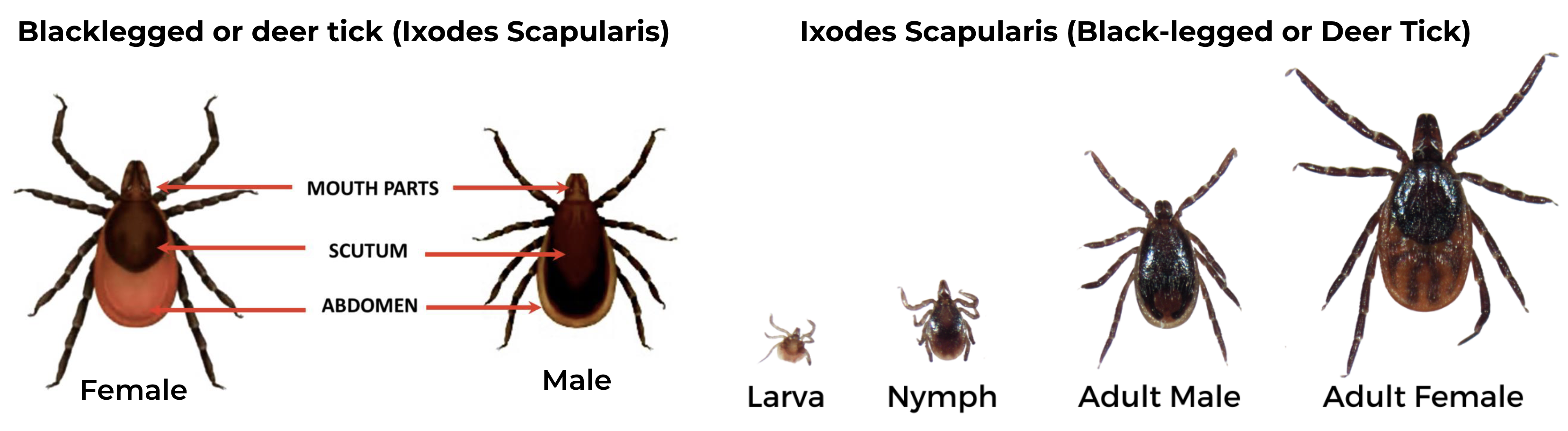
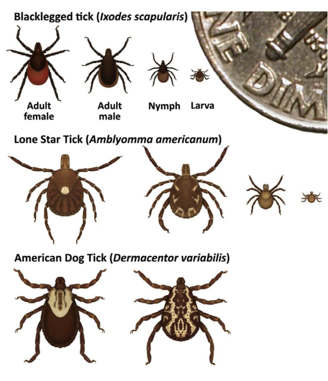
Black-legged Tick (Ixodes scapularis), also known as Deer tick
Geographic Range: Primarily in the northeastern, north-central (Midwest), and southeastern US.

Potential pathogens transmitted:

Female has an orange-brown abdomen, solid black scutum and uniformly black legs. The make has shorter mouthparts, with a scutum that extends over the abdomen

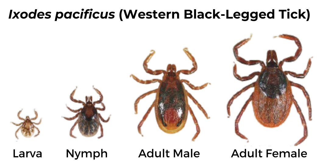
Western Black-legged Tick (Ixodes pacificus)

Geographic Range: Found along the Pacific coast, especially in Northern California.

Pathogens Transmitted:

This tick is identical in appearance to the black-legged or deer tick, but it only occurs in the western coastal states and their ranges do not overlap.

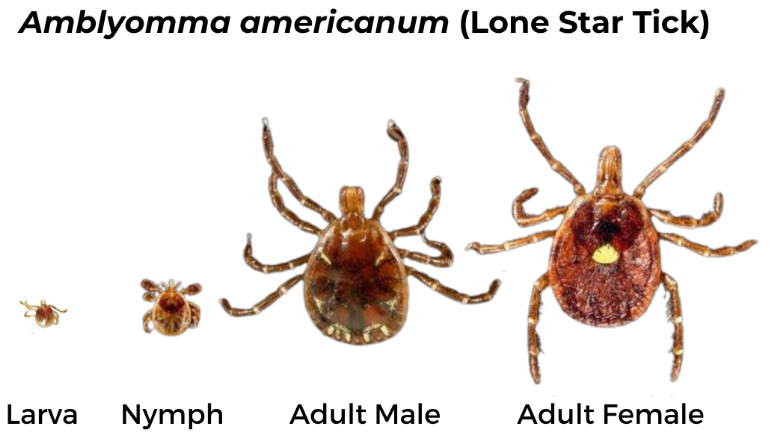
Lone Star Tick (Amblyomma americanum)

Geographic Range: Widely distributed in the southeastern U.S. but has been expanding northward and westward in recent years

Pathogens Transmitted:

*Also associated with alpha-Gal syndrome

Female has long, narrow mouthparts, a bright white mark on the scutum and brown legs with small white bands. The male has long mouthparts, dark brown scutum covering the abdomen and white markings on the end of the abdomen.

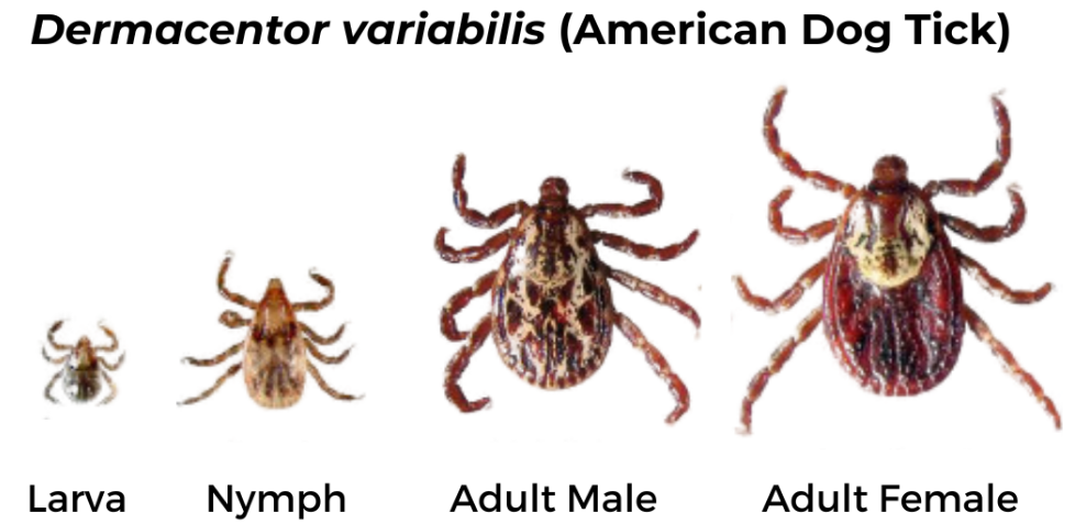
American Dog Tick (Dermacentor variabilis)

Geographic Range: Found east of the Rocky Mountains and in parts of the Pacific Coast.

Pathogens Transmitted:

Female has short mouthparts, white scutum, and brown legs with small white bands. Male has short mouthparts and scutum with white and black markings extending over the abdomen and brown legs with small white bands.

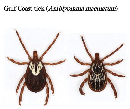
Gulf Coast tick (Amblyomma maculatum)

Female has long mouthparts, white scutum, and brown legs. Male has long mouthparts and black and white scutum covering the abdomen.

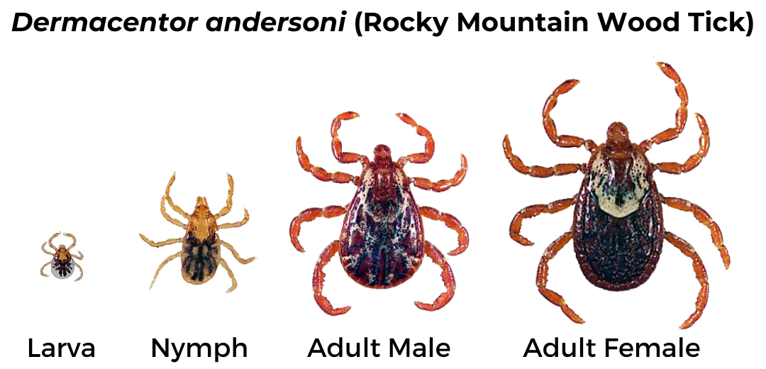
Rocky Mountain Wood Tick (Dermacentor andersoni)

Geographic Range: Found in the Rocky Mountain states and southwestern Canada.

Pathogens Transmitted:

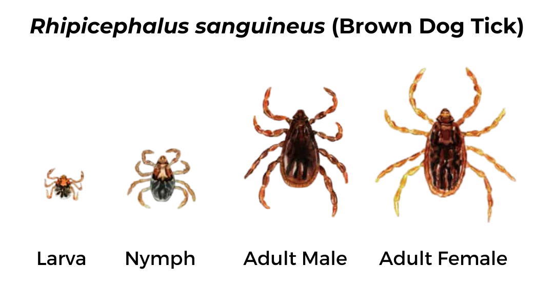
Brown Dog Tick (Rhipicephalus sanguineus)

Geographic Range: Found throughout the U.S.

Pathogens Transmitted:

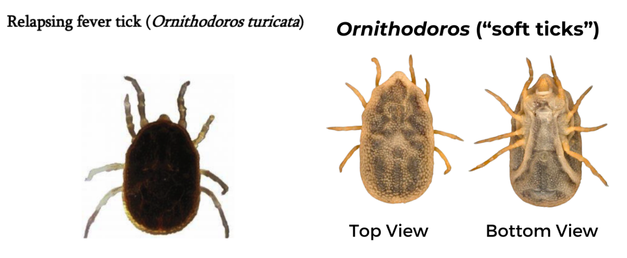
Relapsing Fever Tick (Ornithodoros)

Geographic Range: Specific to each species, but some are found in the western US, typically restricted to caves and burrows, unlike hard ticks which live in grasses.

  • Ornithodoros hermsi tends to be found at higher altitudes (1500 to 8000 feet) where it is associated primarily with ground or tree squirrels and chipmunks.
  • Ornithodoros parkeri occurs at lower altitudes, where they inhabit caves and the burrows of ground squirrels and prairie dogs, as well as those of burrowing owls.
  • Ornithodoros turicata occurs in caves and ground squirrel or prairie dog burrows in the plains regions of the Southwest, feeding off these animals and occasionally burrowing owls or other burrow- or cave-dwelling animals.

Pathogens Transmitted:

Both male and female ticks have similar appearance. Mouthparts are hidden underneath the body and there is no scutum. Body is dark brown and legs are light brown.

LIFE STAGES

Larva and nymph are similar than the adult female, only smaller. Larva has only six legs. All stages will bite humans, except for the larva and nymph of the American dog tick.

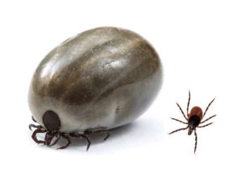
Engorged ticks can obscure mouthparts and scutum.

Should I send tick for testing?

If I find a tick on myself, kids, or pets, should I send it in for testing? Why/why not?
Tick testing is a process in which ticks are tested for the presence of microorganisms that could cause human illnesses such as Lyme disease. It can be useful for tracking the geographic distribution of tick-borne diseases, but it is not a diagnostic test and cannot tell you if you have, or will get a tick-borne illness.

Tick testing is generally unnecessary and should not be a substitute for diagnosis by a physician. Just because a microorganism is found inside a tick does not mean the tick is capable of transmitting that microorganism to a human. Remember: tick physiology is vastly different from humans.

Tick testing is not usually done in a clinical laboratory. Clinical labs must adhere to high quality control standards that do not apply to non-clinical labs. As a result, results may not be accurate and should not guide treatment decisions.

Waiting for tick testing results to make treatment decisions can be risky, especially if symptoms develop before results are available.

Just as a positive test cannot tell you if you’ve actually been infected with a microorganism identified in a tick, a negative test can give false security, as it is possible another tick bit you and did transmit a pathogen.

You can send a tick in for testing if you want, but not doing so does not increase risk of illness. You should be aware of the limitations to the information gained from doing so.

If I send a tick in for testing, does it mean I am infected with whatever was detected?
Positive test results from tick testing does not inherently mean a person was infected, even if results are accurate. Ticks need to attach and feed for specific time periods to be able to transmit pathogens to a human. For example, an infected tick generally has to be attached for at least 36 hours to transmit the bacteria that causes Lyme disease.

In addition, just because a microorganism is found inside a tick does not mean the tick is capable of transmitting it to a human. Remember: tick physiology is vastly different from a human, and a microorganism needs to overcome multiple challenges in order to be successfully transmitted and survive in a different host.

What should I do instead?

If you live where there is high prevalence of Lyme disease, contact your physician (within 72 hours) if you are bitten by a black-legged tick and it has been attached for more than 24 hours. High-risk bites can be treated with prophylactic antibiotics such as a single dose of doxycycline if started within 72 hours after tick removal.

Monitor for symptoms and see a physician if you develop signs of tick-borne illness in the weeks following tick removal or being in tick habitat (forests and fields, especially those with mice and deer) such as: fever, fatigue, chills, aches and pains, or rash.

Ticks can be potential vectors for illness, but it doesn’t mean they automatically are. Being proactive about prevention and prompt removal is your best defense.

Sources:

  • Somewhat Common (more than 1,000 cases per year) 

Rocky Mountain Spotted Fever (RMSF)

Is the most severe tick-borne rickettsial illness in the United States. It is caused by the bacterium Rickettsia rickettsii, an intracellular pathogen that primarily targets the endothelial cells lining the blood vessels, leading to vascular injury.

The primary vectors responsible for the transmission of RMSF are Dermacentor variabilis, commonly known as the American dog tick, Dermacentor andersoni, or the Rocky Mountain wood tick, and Rhipicephalus sanguineus, known as the brown dog tick, which has emerged more recently as a significant vector, especially in parts of the southwestern U.S. and the U.S.-Mexico border region.

There are typically between 250-6,000 cases of tickborne spotted fevers reported in the US every year. Because of genetic similarities of other rickettsial bacteria to Rickettsia rickettsii, it can be difficult to determine which proportion of these cases are specifically RMSF.

While historically associated with the Rocky Mountain region, RMSF cases have been reported throughout the contiguous U.S., with more than 60% of reported RMSF cases occurring in five states (North Carolina, Tennessee, Oklahoma, Arkansas, and Missouri). In recent years, the disease’s range has expanded, especially in parts of the southwestern US.

Symptoms of RMSF typically begin within a week after a tick bite and may be quite severe. They can include high fever, severe headache, muscle pain, chills, fatigue, nausea, vomiting, abdominal pain, and a characteristic red, non-itchy rash that typically appears a few days after the fever (though not all patients develop this rash). The RMSF-associated rash often starts on the wrists, arms, and ankles and can spread to the torso. Severe or untreated illness can lead to organ damage to the heart, lungs, kidneys, and brain, organ failure, and death.

If treated promptly, the mortality rate for RMSF is less than 1%. However, if left untreated, the mortality rate can rise to 20% or higher. The antibiotic doxycycline is the first-line treatment for RMSF in patients of all ages, including children. Early intervention is crucial, as delays in treatment can lead to more severe disease and potential fatalities. Alternative treatments are less effective and can be associated with more severe outcomes.

Diagnosis of RMSF is typically based on clinical presentation and history of potential exposure to tick habitats. Laboratory testing such as serology, PCR, and skin biopsy can be used for confirmation, but due to the rapid progression of RMSF and severity of illness, treatment is typically not delayed while awaiting test results if RMSF is suspected.

Human granulocytic anaplasmosis (HGA)

Is a tick-borne disease caused by Anaplasma phagocytophilum, a rickettsial bacteria [41].  HGA has been gaining increased attention in recent years due to rising incidence and potential for severe health outcomes.

In the United States, it is most common in the Northeast and northern Midwest where the pathogen is transmitted by Ixodes scapularis, but can also be spread by its western counterpart, Ixodes pacificus, found in the Pacific coastal regions of the US.  It overlaps geographically with other diseases transmitted by these ticks, including Lyme disease and babesiosis. The geographic range and number of cases in the United States have increased over the past 20 years from 348 cases in 2000 to 5,655 cases in 2019.

The bacterium that causes HGA, Anaplasma phagocytophilum, infects white blood cells, specifically granulocytes, which play a critical role in the immune response. The infection of these cells is central to the symptoms and complications of the disease.

Clinical manifestations of HGA range from asymptomatic to severe disease and death.  After an incubation period of one to two weeks following a bite from an infected tick, patients most commonly experience flu-like symptoms such as fever, sweats, chills, headache, myalgia, and arthralgia. Additional gastrointestinal symptoms like nausea, vomiting, and diarrhea may also be present. More severe symptoms with respiratory, renal, and neurological complications occur primarily in immunocompromised hosts and can lead to organ failure and death. The overall mortality rate is less than 1%. Rarely, asymptomatically infected patients may transmit the infection by donating blood.

The mortality rate of HGA is less than 1% with appropriate treatment but can be higher in immunocompromised individuals or if left untreated. Over the past several decades, there has been an increase in reported HGA cases in the U.S., coinciding with increased tick prevalence.

Diagnosing HGA can be challenging due to its non-specific symptoms. Laboratory tests play a pivotal role in its confirmation. The diagnosis of HGA is usually confirmed by PCR amplification of A. phagocytophilum DNA  in blood. Infected blood can also occasionally be visualized microscopically. A four-fold rise in A. phagocytophilum antibody titers in acute and convalescent sera can also be used to confirm the diagnosis. A clinical diagnosis based on symptoms and potential tick exposure may warrant starting treatment even before lab results are available.

The treatment of choice is the antibiotic doxycycline which results in rapid improvement and is usually continued for a 10-day course. Other supportive measures might be needed depending on the severity of the patient’s condition. Currently, there’s no vaccine available for HGA, underscoring the importance of preventing tick bites.

Babesiosis

Is an emerging infectious disease caused by protozoan parasites. The causative agents of babesiosis are protozoan parasites from the Babesia genus, with Babesia microti being the most common species affecting humans in the United States. These parasites infect red blood cells, leading to the characteristic symptoms of the disease. 

Babesiosis is usually transmitted by Ixodes scapularis ticks but can also be transmitted through transfusion of blood and blood components, transplantation of solid organs, or by perinatal transmission. In 2019, 2,418 cases of babesiosis were reported in the US, most from the Northeast and the upper Midwest. The most highly endemic states include Connecticut, Massachusetts, Maine, Minnesota, New Jersey, New York, Rhode Island, Vermont, and Wisconsin. The true number of cases may be up to 10-fold higher than the number of reported cases. Less than 25 cases due to infection with Babesia duncani have been reported on the West Coast.

Babesia infection can range from asymptomatic infection to life threatening disease, depending on factors like the individual’s age and immune status. The incubation period after tick bite is 1 to 4 weeks, but it is 1 to 9 weeks (but sometimes as long as 6 months) following transfusion of infected blood or blood components. Babesia infection is asymptomatic in about 20% of immunocompetent adults and 40% of children.

The most common clinical presentation is a mild viral-like illness, with intermittent fever, fatigue and myalgias. Patients with moderate to severe disease experience fever, fatigue, chills, sweats, nausea and anorexia, headache, and myalgia. Illness typically lasts for a week or two. In some instances, hemolytic anemia (anemia caused by rupture of red blood cells) can arise. This occurs because the parasite invades and destroys red blood cells. Immunocompromised patients, including newborn infants; individuals over 50 years of age; those with asplenia, cancer, HIV/AIDS, immunosuppressive therapy, or preexisting cardiac, hematologic, liver, or pulmonary disease; or those who acquired babesiosis through blood transfusion often experience severe acute illness that usually requires hospital admission.

Complications include acute respiratory distress syndrome (ARDS), severe anemia, congestive heart failure, renal failure, disseminated intravascular coagulopathy (DIC), decreased level of consciousness, splenic rupture, and multiorgan failure syndrome [59,60,71-75]. The overall mortality rate for babesiosis is low, but it can reach up to 28% in high-risk individuals, such as the elderly, the immunocompromised, or those without a spleen. Some patients may have symptoms for as long as 27 months despite standard antiparasitic treatments.

Diagnosis should be considered in patients living in or traveling through an endemic area for babesiosis and having typical Babesia symptoms. Babesiosis may also be considered in patients who had a blood transfusion within the past six months, although this is now a rare possibility because of recent widespread screening of the blood supply. The diagnosis is confirmed by identification of parasites on blood smear and/or PCR.

The standard treatment for babesiosis is a combination of two medications: atovaquone and azithromycin, and generally is highly effective. Clindamycin and quinine is an alternative choice, especially in severe cases. Exchange transfusion can also be used in severe cases. Prolonged therapy for at least 6 weeks is recommended for severely immunocompromised patients who do not clear parasitemia and improve symptomatically after 7 to 10 days of therapy.

Ehrlichiosis

Is caused by a bacterial infection caused by species of Ehrlichia in the US. Ehrlichia chaffeensis and Ehrlichia ewingii can be transmitted to humans through the bite of the lone star tick (Amblyomma americanum). Less commonly, E. muris eauclairensis has been found to cause Ehrlichiosis after transmission by the bite of the black-legged tick (Ixodes scapularis). These tick species are also capable of transmitting other pathogens, underscoring the importance of these vectors in public health. E. chaffeensis causes the majority of cases reported in the US.

While cases of ehrlichiosis have been reported across the U.S., its primary range aligns with that of the lone star tick. Ehrlichiosis is most common in the southeastern US, and can extend into parts of the Midwest and the eastern states. As tick populations and their ranges shift, the geographic range of ehrlichiosis may also change. Ehrlichiosis is relatively uncommon: 2,093 cases were reported in the US in 2019, with over 50% of cases occurring in 4 states: Missouri, Arkansas, North Carolina, and New York.

Symptoms of ehrlichiosis usually begin 1-2 weeks after a tick bite and are generally non-specific. They include high fever, fatigue, headache, muscle aches, chills, and nausea and/or vomiting. In about 33% of cases, a rash is reported, and more often occurs in children than adults. The rash develops around 5 days after fever begins, and appears as red splotches or pinpoint dots. More severe cases can involve neurological manifestations such as menigoencephalitis, respiratory failure, hemorrhage, and multi-organ failure. These severe symptoms are more common in individuals at high risk, such as those who are immunocompromised or very young or elderly. The estimated mortality rate is around 1-3% and is higher among high risk individuals.

Diagnosing ehrlichiosis can be challenging due to its non-specific symptoms. A combination of clinical signs, history of tick exposure, and laboratory testing aids diagnosis. Lab tests include blood smears, serology, and PCR. However, treatment should commence if ehrlichiosis is suspected, even before lab confirmation, given the potential severity of the disease.

The antibiotic doxycycline is the preferred treatment for ehrlichiosis in both adults and children of all ages. Prompt treatment Starting treatment early is crucial to prevent more severe complications and to speed recovery.

  • Uncommon or rare:

Powassan virus (POWV)

Also called deer-tick virus, has emerged as a public health concern, especially in regions where Lyme disease is endemic. The Powassan virus is a member of the Flaviviridae family and is closely related to tick-borne encephalitis in northern Eurasia and more distantly related to other well-known viruses like the West Nile, Dengue, and Zika viruses. POWV is an RNA virus, which means that its genetic material is made of ribonucleic acid rather than deoxyribonucleic acid (DNA). This RNA structure can lead to higher mutation rates, potentially influencing the virus’s virulence and transmission dynamics.

POWV was first discovered in 1958 from a fatal case occurring in Powassan, Ontario and named after that location. This incidence case was caused by the bite of an Ixodes cookei tick, also known as the groundhog tick, which normally feeds upon wildlife (skunks, weasels, groundhogs), only rarely biting humans. Subsequent cases were also rare, less than one per year, for decades after its initial discovery.

In 1997, Powassan virus was found in the black-legged or deer tick Ixodes scapularis, which more commonly feeds on humans   and the prevalence of infection in this tick has increased dramatically since then, as has the incidence of human infection. Over 200 cases have been reported to the CDC over the last decade. In the United States, cases have predominantly been reported in the Great Lakes region (upper Midwest) and the Northeast. However, as tick populations spread, the range of POWV could potentially expand. Unlike other pathogens transmitted by Ixodes scapularis, Powassan virus can be transmitted within minutes, not hours, after a tick bite and all stages (larvae, nymphs and adults) can be infected. Contact your local or state health department to find out if cases have been reported in your area.

Fortunately, most individuals infected with POWV may remain asymptomatic. However, if symptoms do develop, they can appear within one week to one month after a tick bite.

Initial symptoms include fever, headache, nausea and general weakness. As infection progresses, severe neurological symptoms can arise, such as confusion, seizures, and more severe cases can progress to meningitis (inflammation of the brain lining) or encephalitis (inflammation of the brain). Given the virus’s potential impact on the central nervous system, long-term effects or disabilities can result, even if the acute phase of the illness is survived.

Death occurs in about 10% of severe cases and approximately half of the people who survive severe disease has long-term health problems such as recurring headaches, loss of muscle

mass and strength, and memory problems.

There is no vaccine to prevent POWV infection and there are no specific antiviral treatments available to cure infection. Treatment of illness is largely supportive, addressing the symptoms rather than the virus itself. This might include hospitalization, respiratory support, and intravenous fluids. Given the absence of a targeted treatment and the potential severity of symptoms, preventive measures such as tick avoidance become crucial.

The recent increase of Powassan virus poses significant health risks due to the mortality rate and potential for severe neurological complications. While the overall prevalence of POWV infections in the U.S. is low, an increase in diagnosed cases over the past few years has raised concerns among health professionals. With expansion of tick populations and the increasing incidence of tick-borne diseases, public awareness and preventive measures are critical. Continued research into targeted treatments and potential vaccines will be necessary to manage this potentially dangerous pathogen.

Borrelia miyamotoi disease (BMD)

Also known as hard-tick relapsing fever, is caused by a spirochete bacterium related to the Lyme disease bacterium Borrelia burgdorferi, but it is in the relapsing fever group of spirochetes rather than in the Lyme disease or borreliosis group. It was originally found from a tick in Japan in 1991 and was found in Ixodes scapularis in Connecticut in 2001. Clinical cases of BMD in the US were not described until 2013. Borrelia miyamotoi has been found in 2% of I. scapularis ticks collected throughout its northern range from New England to the Mid-West. It has also been found in Ixodes pacificus in the western states.

Borrelia miyamotoi infection in ticks can be passed from an adult female tick to offspring through infected eggs. Thus, all stages (larva, nymph and adult) can transmit the infection to humans by tick bite. The time required for transmission of spirochetes by tick bite may be as soon as 24 hrs, with increasing probability of 75% after complete feeding.

Although not a mandatory reportable disease, more than 100 cases per year are estimated to occur. This is surely an underestimate considering its wide geographic distribution, prevalence in ticks, mode of transmission in ticks, and efficient transmission from tick to humans. BMD is considered to be an emerging disease by the CDC.

Symptoms of BMD include flu-like symptoms: fever, fatigue, sleepiness, chills, muscle and joint stiffness, aches and pains, and nausea. BMD does not cause the EM skin rash associated with Lyme disease. Like other relapsing fever diseases, symptoms may reappear after a period without symptoms. Infection in immunocompromised patients can be much more severe. In combination with generalized flu-like symptoms, immunocompromised patients often exhibit reduced cognition, disturbed gait, memory deficits, confusion, and other neurological deficiencies resulting from inflammation of the brain. Coinfection with B. burgdorferi is possible, but no cases have yet been reported.

Diagnosis of BMD can be difficult. There is currently no specific standardized diagnostic test approved by the FDA. Spirochetes can be visualized in peripheral blood or detected in blood by PCR. Despite its relationship to B. burdgorferi, it cannot be detected by standard antibody tests for Lyme disease. Except for an EM rash, the symptoms are similar to Lyme disease, but symptoms of Lyme disease do not relapse.

Treatment for BMD typically follows the guidelines used for Lyme disease. The antibiotics doxycycline, amoxycillin, and ceftriaxone have been successful in clearing infection. Thus, patients infected with B. miyamotoi, misdiagnosed as Lyme disease, would benefit from the same treatment.

Tularemia

Colloquially known as “rabbit fever,” is a bacterial infection caused by Francisella tularensis. Francisella tularensis is one of the most infectious pathogenic bacteria known; as few as 10 organisms can cause disease in humans, which is an exceedingly low infectious dose.

 While not only transmitted by ticks, F. tularensis can be transmitted by the wood tick (Dermacentor andersoni), the lone star tick (Amblyomma americanum), and the dog tick (Dermacentor variabilis). F. tularensis can also infect humans through multiple routes of exposure, including inhalation, ingestion, and direct skin contact, and other arthropod bites. The bacterium can also survive for weeks in the environment, especially in water and soil. This persistence facilitates its transmission through various means, such as contaminated water or aerosols. This versatility makes it a significant concern from both natural outbreaks and potential bioterrorism perspectives.

Tularemia can be found throughout the U.S., but it’s more common in the south-central, Pacific, and western regions. Outside of the U.S., it has been reported in various parts of the world, especially in the northern hemisphere. While not as prevalent as other zoonotic diseases, several hundred cases are reported annually in the US.. 270 cases of tularemia were reported in 2019.

Symptoms of tularemia depend on the route of infection, with the disease presenting in various forms. After a tick bite, the following forms of tularemia can present:

  • Ulceroglandular Tularemia: This is the most common form of tularemia following a tick bite. Patients with this form often develop a skin ulcer at the site of the tick bite. The ulcer is accompanied by swelling of regional lymph nodes, usually those that are nearest to the site of the tick bite.
  • Glandular Tularemia: Similar to ulceroglandular tularemia, this form involves swollen lymph nodes but without the presence of an ulcer. It’s less common than the ulceroglandular form but can also result from a tick bite.
  • Oculoglandular Tularemia: Though more commonly associated with direct contamination of the eye (e.g., from handling infected animals and then touching the eye), it can occasionally present after a tick bite, especially if one touches the bite and then their eye. It’s characterized by eye inflammation, pain, and discharge, along with swollen lymph nodes.
  • Typhoidal Tularemia: This is a more generalized form of the disease, presenting without localized symptoms. It can be a result of any route of infection, including tick bites. Patients might experience fever, fatigue, and other systemic symptoms without obvious ulcers or lymph node involvement. This form can be particularly severe and can sometimes be mistaken for other febrile illnesses.
  • Pneumonic Tularemia: While this form is most often associated with inhalation of the bacterium, it can also arise secondarily from other forms of tularemia, potentially including those initiated by a tick bite. Pneumonic tularemia involves cough, chest pain, and difficulty breathing.

One other presentation of tularemia, oropharyngeal tularemia, can be acquired from consuming contaminated food or water. Symptoms impact the mouth, throat, and digestive system and include sore throat, lymphadenopathy, mouth ulcers, tonsillitis, pharyngitis, abdominal pain, diarrhea, vomiting, and fever.

With appropriate treatment, the mortality rate for tularemia is around 1%. However, certain forms, especially pneumonic and typhoidal, can have mortality rates up to 7% if untreated.

Tularemia’s diverse clinical presentation can make initial diagnosis challenging. However, a combination of clinical presentation, history of potential exposure (like tick bites or handling wildlife), and laboratory tests can confirm the diagnosis. Serologic tests, cultures, and PCR are commonly used, though biocontainment procedures must be implemented in handling patient samples due to the bacterium’s high infectivity.

The antibiotic streptomycin has traditionally been the drug of choice for treating tularemia, but gentamicin can also be effective. In mild cases or for patients allergic to aminoglycosides, the antibiotic doxycycline might be used.

Rickettsia parkeri Rickettsiosis

Is an emerging tick-borne disease caused by the rickettsial bacterium Rickettsia parkeri, a relative of Rickettsia rickettsii, the bacterium that causes Rocky Mountain Spotted Fever. Rickettsia parkeri is a member of the spotted fever group of Rickettsia, a collection of bacteria that cause similar clinical symptoms but vary in disease severity and specific presentations.

Rickettsia parkeri is transmitted by the Gulf Coast tick, Amblyomma maculatum. Rickettsia parkeri infection is rare with only around 40 cases have been reported since the illness was defined in 2004. Historically, Rickettsia parkeri Rickettsiosis has been associated with the southeastern and south-central regions of the U.S., in line with the geographic range of the Gulf Coast tick. However, cases have now been reported in several states, expanding the known range of this disease.

Symptoms of R. parkeri infection typically appear within 2-10 days after a tick bite and often have overlap with other rickettsial spotted fevers, including fever, fatigue, headache, muscle aches, a distinctive skin rash. Lymphadenopathy (swollen lymph nodes) may also present near the site of the tick bite. In addition, an eschar (a dark, scab-like lesion resembling a sore or a pimple) can be found at the site of the tick bite, distinguishing R. parkeri rickettsiosis from some other rickettsial diseases, and is often the first symptom of infection.

Rickettsia parkeri Rickettsiosis is generally considered milder compared to other rickettsial diseases like Rocky Mountain spotted fever. No identified fatalities have been reported in the US to date, and many patients recover without complications. The antibiotic doxycycline is the recommended treatment for all age groups.

 Like other rickettsial spotted fevers, Rickettsia parkeri Rickettsiosis is diagnosed using a combination of clinical symptoms, history of tick exposure, and laboratory testing. Serologic tests can detect antibodies to R. parkeri, while PCR and culture can identify the bacterium in clinical samples. The presence of an eschar can be a helpful diagnostic clue, but not all patients will have this manifestation.

Tick-borne Relapsing Fever (TBRF)

Is caused by infection by one of several different species of spirochete bacteria of the genus Borrelia. These constitute a group of bacteria that is distinct from the group of spirochetes that cause Lyme disease. In western and central North America the two species that cause infections of humans are Borrelia hermsii and Borrelia turicatae. Other species of Borrelia cause TBRF in South America, Africa, and Eurasia. Another RF agent is Borrelia recurrentis, which is transmitted by the human body louse and not by ticks. Louse-borne relapsing fever has caused large epidemics in the past in which there is spread between people via lice. Louse-borne relapsing fever continues to occur in parts of Africa but not elsewhere. (Borrelia miyamotoi is also a member of the RF group of species but is transmitted is transmitted by hard ticks of the genus Ixodes.)

These bacteria are transmitted to humans through the bite of soft ticks of the genus Ornithodoros. Soft ticks differ in two important ways from the more familiar “hard ticks” that can cause other tick borne illnesses in humans (e.g., the dog tick and the deer tick). First, unlike the more commonly known hard ticks, soft ticks tend to feed quickly, with feeding lasting less than half an hour. Soft ticks often bite hosts during the night and then retreat to their natural habitats, making them less noticeable. Second, soft ticks do not search for prey in tall grass or brush and they do not quest like hard-bodied ticks. Instead, they tend to live within nests in burrows, logs, rock piles, caves, or within walls and ceilings of cabins and houses, feeding as needed on the rodent or other mammal as it sleeps.

The unique feeding and nesting behavior of soft ticks dictates the risk of disease aquisition. Borrelia hermsii, spread by Ornithodoros hermsi ticks, is overall the most commonly reported cause of TBRF in the US. Infection with B. hermsii is commonly associated with rustic cabins where rodents, such as chipmunks, build nests within wall and ceiling spaces. Its distribution ranges from foothills to high elevations of mountainous regions of western North America. Borrelia turicatae, transmitted by Ornithodoros turicata, is typically found at lower elevations of more arid areas of western North America. B. turicatae acquisition is often associated with entry into or sleeping in caves that provide shelter for a variety of small and large mammals.

The incidence of TBRF in the U.S. is low, with 251 total cases reported between 2012-2021, occurring in 11 states, with median case prevalence of 24 reported cases of TBRF every year. Over 70% of cases occur in 3 states: California, Washington, and Colorado. But it is likely under-reported because of the non-specific nature of the presenting illness, lack of diagnostic capacity in most healthcare facilitiies, and empirical use of antibiotics of undifferentiated febrile illnesses by healthcare practitioners.

TBRF has a very distinctive clinical presentation, where patients experience two or more episodes of fever separated by intervals of apparent recovery. These recurrent episodes of fever typically last 2-7 days, separated by afebrile periods of 3-7 days. The first episode may be diagnosed as a viral illness and managed only with symptomatic treatment. Not uncommonly the diagnosis is made when the patient returns to emergency department or clinic with a return of the fever. Other nonspecific symptoms can also be present including headache, muscle and joint aches, chills, and nausea. The name “relapsing fever” derives from these cyclical fever episodes that can recur several times before illness resolves in untreated patients. The bacteria in each relapse are variants that are able to escape from the antibodies that first appear in response to the initial infection.

During each febrile period the temperature may reach 103-104 degrees F. and commonly is accompanied by shaking chills. The febrile period ends over a few to several hours and is accompanied by intense sweating as the body temperature declines. During this time the patient may also have marked fatigue and low blood pressure that calls for intravenous fluids and other support in the hospital. Some patients, especially those infected with B. turicatae, experience neurologic involvement, including facial muscle weakness (Bell’s palsy) or a type of meningitis.   

While TBRF can be debilitating during febrile periods, it’s generally not fatal, especially with timely treatment and if there is not underlying immune deficiency or malnutrition. The mortality rate is estimated to be less than 2% globally, and death due to TBRF is rare in the US. Many milder cases of TBRF may go unnoticed or misdiagnosed as anacute viral illness”, and an individual’s immune system eventually clears infection after a few cycles of illness even without treatment.

Findings on physical exam vary depending on the severity of illness and when the patient seeks medical care. Regardless, there are no findings, such as a particular type of rash, specific for TBRF. There may be enlargement of the spleen.

Diagnosing TBRF involves a combination of clinical presentation, history of potential exposure, and laboratory testing. The spiral-shaped bacterium can often be seen in smears of blood taken at the time of the fever. The smears are processed with Wright or Giemsa staining as for routine differential counts of white cells by microscopy. Between febrile episodes, the bacteria cannot be seen in blood smears. PCR assays of the blood for RF species are available from some commercial reference laboratories. Tests of antibodies to RF Borrelia species are available from some laboratories but are only useful for retrospective confirmation of the diagnosis after the patient has recovered.

RF is treated with antibiotics. This is usually a penicillin or cephalosporin given intravenously or doxycycline, a tetracycline, given orally. An alternative to the tetracycline for pregnant women is erythromycin or related macrolide antibiotic. With any antibiotic treatment there commonly may be a temporary worsening of the clinical status within an hour or two of administration of the first dose. This includes a rise in temperature and a fall in blood pressure. This phenomenon is called a Jarisch-Herxheimer reaction, and it is in response to the death of many bacteria all at once. Recovered patients may experience some post-infection fatigue, but there is no evidence of persistent infections with a TBRF spirochete either after antibiotic therapy or a natural cure.

There is no vaccine to prevent TBRF. If there has been known exposure to RF, such as an overnight stay in a cabin where there has been another case, a single or a few doses of doxycycline can prevent infection.

Colorado Tick Fever (CTF)

Is one of a few tick-borne illnesses in the United States caused by a viral pathogen, specifically, the Colorado Tick Fever Virus (CTFV). CTF is quite rare, with only 59 cases reported between 2010 and 2019. CTFV is a virus in the Coltivirus genus, and is somewhat related to a similar tick-borne virus in Europe, Eyach virus, which can also cause illness in humans. CTFV is an RNA virus, which means that its genetic material is made of ribonucleic acid rather than deoxyribonucleic acid (DNA). CTFV was first isolated from human blood in 1944.

CTFV is transmitted to humans through the bite of the Rocky Mountain wood tick, Dermacentor andersoni, and cases of CTF are almost exclusively restricted to higher elevations (altitudes from 4,000 to 10,000 feet) in the Rocky Mountain region of the US. As of 2020, CTF is a locally notifiable disease in nine states: Arizona, Colorado, Idaho, Montana, New Mexico, Oregon, South Dakota, Utah, and Wyoming.

Symptoms of CTF typically appear between 1 and 14 days following an infected tick bite. Initial symptoms most commonly include fever, chills, headaches, muscle pain, and malaise. Less common symptoms can include pain behind the eyes, photophobia (light sensitivity), abdominal pain, hepatosplenomegaly, nausea and vomiting, sore throat, and rarely, a flat or pimply rash. CTFV often infects erythrocytes (red blood cells), which can lead to biphasic illness in about half of affected individuals. This means symptoms will often dissipate for a few days and then reappear again.

Most cases of CTF are mild, and illness is self-limiting. Affected individuals generally recover completely, although post-illness fatigue and weakness (post-infection sequelae) may persist for several weeks. In rare instances CTF can progress to more severe illness involving the central nervous system and lead to complications such as aseptic meningitis (presenting with stiff neck) and encephalitis (presenting with confusion). Death due to CTF is exceedingly rare.

Diagnosis is primarily based on clinical symptoms and a history of potential exposure to the vector tick in endemic areas. Laboratory tests, such as serology and PCR, can be used to confirm the diagnosis. Given the overlapping symptoms with other illnesses, differential diagnosis is crucial to rule out other tick-borne diseases or conditions that might present similarly.

There is no vaccine to prevent CTFV infection and there are no specific antiviral treatments available to cure infection. Treatment of illness is largely supportive, addressing the symptoms rather than the virus itself. This typically includes rest, fluids, and over-the-counter analgesic and antipyretic medications. In rare cases of severe CTF, some individuals may be hospitalized for intravenous fluids and medications.

Heartland Virus Disease (HVD)

Is a rare but emerging tick-borne infection caused by the Heartland virus (officially Heartland bandavirus) (HRTV). HRTV is a Phlebovirus in the Bunyavirus order, which are segmented genome RNA viruses. Distant relatives include the more commonly known Hantaviruses and Arenaviruses (Lassa virus, Junin virus). HRTV was first discovered in 2009 in Missouri by Dr. Scott Folk of Heartland Regional Medical Center.

HRTV is transmitted through the bite of infected Lone star ticks (Amblyomma americanum), which is also the vector capable of spreading Rocky Mountain Spotted Fever, Ehrlichiosis, Tularemia, southern tick-associated rash illness (STARI), and Bourbon Virus Disease.

Currently, HRTV is quite rare, but cases of the Heartland virus have been recognized in several states, including Missouri, Arkansas, Illinois, Indiana, and Oklahoma. It is likely the range of HRTV overlaps with the endemic range of the Lone star tick. As of November 2022, 60 cases of HVD have been reported.

Symptoms typically present between a few days to 2 weeks following a tick bite. Signs and symptoms are similar to several other tick-borne illnesses including ehrlichiosis and include fever and chills, fatigue, headache, muscle/joint pain, decreased appetite, nausea, and diarrhea. Some infected individuals may have low white blood cell counts (leukopenia) and low platelet count (thrombocytopenia). HVD can become quite severe, especially if immune cell function is impacted. While identified cases are rare to date, many have required hospitalization. Current reported mortality rate, based on the limited number of identified cases, is around 10%, but this is likely an overestimation as most unreported cases were likely mild. As such, it’s important to recognize that the true prevalence and mortality rate may differ as more cases are identified and studied.

The diagnostic process for the Heartland virus remains challenging. Given its recent emergence, standard diagnostic tests are not widely available. Typically, diagnosis hinges on a combination of clinical presentation, history of tick exposure, and specialized laboratory tests, often in collaboration with health departments or the CDC. Clinical criteria have recently been defined, which include: fever greater than 38 °C (or 100.4 °F). thrombocytopenia or platelet count lower than 150,000 per mm3, leukopenia or white blood cell count lower than 4,500 cells per mm3, and evidence of a recent infection with Heartland virus (detection of viral RNA in the blood or tissues by reverse transcription polymerase chain reaction, or by viral neutralization assays using convalescent serum from the patient).

There is no vaccine to prevent HRTV infection and there are no specific antiviral treatments available to cure infection. Treatment of illness is largely supportive, addressing the symptoms rather than the virus itself. This typically includes rest, fluids, and over-the-counter analgesic and antipyretic medications. In rare cases of severe HVD, some individuals may be hospitalized for intravenous fluids and medications.

Bourbon Virus Disease (HVD)

Is a rare but emerging tick-borne infection caused by the Bourbon virus. BVD is a Thogtovirus in the Orthomyxovirus family, which are segmented genome RNA viruses. Bourbon virus was first identified in 2014 in a man in Bourbon County Missouri and isolated by CDC researchers from those patient blood samples.

Thogtoviruses almost never cause illness in humans. The case in 2014 was only the eighth human illness caused by a thogtovirus, and there have been only 5 cases of Bourbon virus reported to date.

The precise vector that can transmit Bourbon virus is still under investigation. However, based on the fact that other Thogtoviruses are transmitted through arthropod bites and the geography and epidemiology of the limited cases of Bourbon viruses in the United States, the leading candidate is the lone star tick (Amblyomma americanum).

Scientists are still learning about possible symptoms caused by this relatively new virus. Symptoms seem to appear about a week after a tick bite, and seem to include fever, fatigue, rash, headache, other body aches, nausea, and vomiting. They also had lower than normal counts of white blood cells (leukopenia) and lower than normal counts of platelets (thrombocytopenia).

Given the limited number of reported cases, determining a precise mortality rate for the Bourbon virus is challenging. However, it is known that the disease can be severe, with some cases being fatal, including the reference case in 2014. Since its discovery, only a handful of cases have been reported, making it a rare but concerning pathogen. Yet, underreporting or misdiagnosis could mean the actual prevalence is higher than currently recognized 

Diagnosing Bourbon virus is complex due to its similarity in presentation to other tick-borne diseases. Currently, there isn’t a widely available standard test for the virus. Diagnosis usually requires specialized laboratory techniques, often involving collaboration with specialized institutions or the CDC.

There are no vaccines or specific antivirals to treat Bourbon virus infection. Care is mainly supportive, focusing on treating symptoms and complications. Hospitalization may be necessary, especially in severe cases or for individuals with other underlying health conditions.

Southern Tick-Associated Rash Illness (STARI)

Is a recent emerging illness that the causative agent has not yet been identified. While its clinical presentation is reminiscent of Lyme disease, caused by the Borrelia burgdorferi bacterium, repeated studies have not conclusively linked this or any other specific pathogen to STARI.

 STARI is believed to be transmitted primarily through the bite of the lone star tick (Amblyomma americanum) due to current epidemiology and geographic range of cases. As the name suggests, STARI is primarily observed in the southeastern and south-central United States, which corresponds to the habitat range of the lone star tick. This includes states such as Missouri, Oklahoma, North Carolina, and parts of Texas, among others.

The symptoms typically manifest within a week of the tick bite and can vary in severity. They can include an expanding “bull’s-eye” Erythema migrans rash similar to that observed in Lyme disease, fatigue, fever, headache, muscle and joint pains, and lymphadenopathy (swollen lymph nodes). Other symptoms associated with Lyme such as asymmetrical arthritis, neurological symptoms, or other persistent symptoms.  

They are key clinical distinctions between STARI and Lyme disease. Patients diagnosed with STARI were more likely to recall a tick bite than were patients with Lyme disease and time interval between tick bite to skin rash was shorter among patients with STARI (6 days compared to an average interval of 2 weeks for Lyme). STARI patients were less likely to have other symptoms in addition to EM, it was less likely to present with multiple EM, and size of EM is smaller and more circular (6-10 cm). While causative agent has not yet been identified, antibiotic treatment with doxycycline has typically been used due to symptom similarity to Lyme. After antibiotic treatment, STARI patients recovered more rapidly than did Lyme disease patients.

STARI is typically not associated with long-term complications and is not known to be fatal. The precise prevalence is difficult to pin down, given the challenges in differentiating it from Lyme disease based on clinical presentation alone. However, it’s believed to be a relatively uncommon condition. Exact annual case numbers are not well-established due to the diagnostic ambiguities and potential underreporting.

Diagnosing STARI is challenging because of its clinical similarities to Lyme disease and the absence of a specific diagnostic test. Typically, diagnosis is made based on clinical presentation, history of tick bite, and exclusion of Lyme disease, especially in areas where Lyme is less prevalent. Laboratory tests currently cannot reliably distinguish STARI from Lyme disease.

Even though the exact causative agent of STARI remains unidentified, treatment with oral antibiotics, particularly doxycycline, has been shown to be effective in alleviating symptoms. It’s believed that early treatment can prevent potential complications or the progression of symptoms.

Alpha-gal Syndrome (AGS)

Is a condition that can be initiated by a tick bite, but is not caused by infection with a pathogen. It is the development of an allergy against a specific sugar found in mammals called alpha-galactosidase (galactose-α-1,3-galactose) or alpha-Gal, which humans don’t produce. The subsequent allergy is called alpha-Gal syndrome (AGS).

Ticks also don’t produce alpha-Gal, but non-human mammals that they feed on do. The primary vector associated with the development of AGS in the U.S. is the lone star tick (Amblyomma americanum). However, other tick species worldwide, such as the Australian paralysis tick and European castor bean tick, have also been implicated in the condition. 

The distribution of AGS correlates with the range of the responsible tick species. In the U.S., this primarily encompasses the southeastern and central states, where the lone star tick is prevalent. However, with other tick species implicated internationally, cases have also been reported in Europe, Australia, and parts of Asia.

 AGS can develop independently of a tick bite, but it is thought that exposure to alpha-Gal through tick saliva can trigger this. It is not a common allergy; currently it is estimated around 1% of the US population have IgE antibodies against alpha-Gal.

In the context of tick-bite associated AGS, it is hypothesized that alpha-Gal molecules from a previous blood meal a tick has taken can end up in the tick’s body and be transferred to a new animal once mixed with the tick saliva. In some humans, this sugar may be recognized as foreign, which triggers an immune response. This is the same process that occurs in the development of other food and environmental allergens.

Individuals with AGS may develop symptoms 2-6 hours following consumption of animal products that contain alpha-Gal. Symptoms are similar to other allergies, and can range from mild to severe and include urticaria (hives), itchy skin, swelling, often around the eyes, lips, or inside the mouth, abdominal pain or cramps, nausea or vomiting, diarrhea, runny or stuffy nose, asthmatic symptoms, and rarely, anaphylaxis, a severe allergic reaction that can be life-threatening.

Diagnosis of AGS involves a combination of patient history and specific blood tests that measure IgE antibodies against the alpha-gal molecule. The delayed onset of symptoms after eating red meat, combined with a history of tick bites, often provides clinical clues.

Management includes avoiding foods that contain this sugar. Fish, poultry, and produce do not contain alpha-Gal. Some medications and medical devices may be derived from animal products containing alpha-Gal so ingredient labels may be important for those with AGS.

There is no cure, but recommendations in the US include reducing risk of bites from the Lone Star tick, which has been linked to AGS. For inadvertent exposures or reactions, antihistamines can be used to treat mild symptoms. In cases of severe reactions, like anaphylaxis, injectable epinephrine is necessary, and individuals diagnosed with AGS are often prescribed an epinephrine auto-injector to carry with them. There is currently no cure for AGS, but some individuals have reported a reduction or resolution of their allergy over time, particularly if they avoid further tick bites.